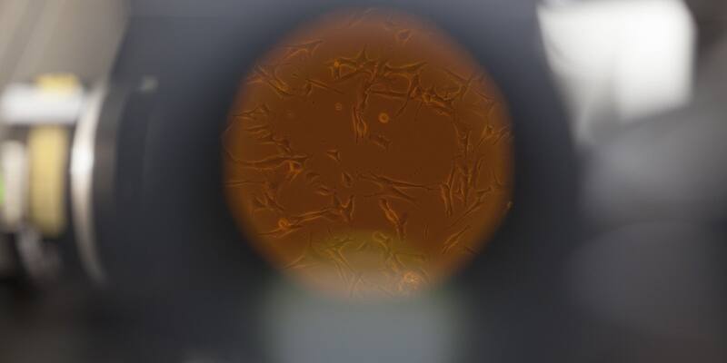
Cells seen through a laboratory microscope at NASA's Kennedy Space Center in 2014. Illustrative photo, not from the Nikon contest. Image: NASA/Dimitri Gerondidakis / Wikimedia Commons, Public domain, cropped
Nikon has disqualified the winner of its 2026 Small World in Motion microscopy video contest, ruling that the clip of beating airway cilia "did not comply with the competition rules regarding generative AI". The decision, posted on October 9, comes three and a half weeks after the video was named the best of the year and about a week after scientists' complaints pushed Nikon into a second review.
The rankings have been reshuffled. Nguyen Nam Nhat of Vietnam, whose clip of a tiny roundworm and a single-celled Dileptus came second, is now the winner, with Benedikt Pleyer's jellyfish larvae in water droplets second and Dr Andrew Moore of the Howard Hughes Medical Institute's Janelia Research Campus third.
What Nikon decided
Nikon said it reached its verdict "after thoroughly re-evaluating the video and supporting materials and consulting with members of our judging panel". It stressed that the ruling was "based solely on the submitted video's eligibility" and "should not be interpreted as a judgment of the entrant's professional reputation, scientific contributions, or intent".
The company also admitted the episode exposed gaps in its own process. Advances in imaging and AI are creating "new challenges for organizations across many fields", it wrote, and the case "has shown the need" for both of its competitions to revisit their rules and evaluation procedures for future entries.
A video scientists said could not be real
The disqualified entry came from Dr Ning Xu, an optical engineer whom the BBC describes as being at Tsinghua University in China and Nature places at the National University of Singapore. Named the winner on September 15, it showed hair-like cilia moving in the airway of a child with primary ciliary dyskinesia (PCD), a rare genetic disorder that causes chronic lung, sinus and ear infections. In the clip the cilia sit on top of red, purple and blue structures that the competition's own write-up did not explain.
Microscopists began picking at it within days. Edward Phelps, a bioengineering researcher at the University of Florida, wrote on LinkedIn that "the purple structures resemble mitochondria but such a sub-epithelial structure composed of extracellular mitochondria of the same size as cell nuclei does not occur in biology". Ian Donovan, a PhD student at UT Southwestern Medical Center, pointed out that the video carried Google's invisible SynthID watermark, the tag Google's AI tools stamp on what they generate and that anyone can now check with its public SynthID Detector.
On September 22, Nikon quietly updated its page about the entry to say that an "unsupervised" AI model had assisted with "post-processing". At first the company said it saw no rule-breaking, before announcing a fuller review.
Dr Robert Hirst of the University of Leicester, lead scientist at the NHS centre for PCD diagnosis and among the first to raise concerns, told the BBC he had spent 20 years diagnosing the condition by studying cilia, and that the cells and cilia in the video "look nothing like those from PCD patients":
I knew immediately that the video was fabricated.
Dr Robert Hirst, University of Leicester, to the BBC
Xu says AI only coloured real footage
Xu has not denied using AI, but says he stayed within the rules. On LinkedIn he wrote that AI was not used to generate the experimental movie, the cilia or their motion. "It was applied afterwards to the reconstructed grayscale data to distinguish and color structures with similar morphology," he said, adding that his team enhanced the regions below the cilia "without making anatomical claims about what those rendered features represent". He also said he had cooperated with Nikon's review and supplied technical documentation. He has not yet responded publicly to the ruling.
"They are data"
Researchers say the problem is not AI in microscopy as such. Markus Sauer, a super-resolution microscopy specialist at the University of Würzburg, told Nature that AI visualisation becomes an issue when it misrepresents, exaggerates or alters what the experiment actually found. Melanie White, a developmental biologist at the University of Queensland, put it more bluntly:
Scientific images are not just illustrations. They are data, and we need to be able to trust that what we are seeing is grounded in the underlying measurement.
Melanie White, University of Queensland, to Nature
It is the second time in a week that AI's place in published science has been questioned, after OpenAI pulled three of the 722 AI-written maths papers it had posted on GitHub.
Why it matters
Small World is one of the best-known showcases for scientific imaging, and its top prize went to a video that experts say showed structures that do not exist. If a specialist jury can miss AI-invented detail, journals and grant panels face the same risk, and a watermark check is fast becoming part of reviewing a scientific image.
Sources: Nikon Small World, Nature, BBC
Latest Business news